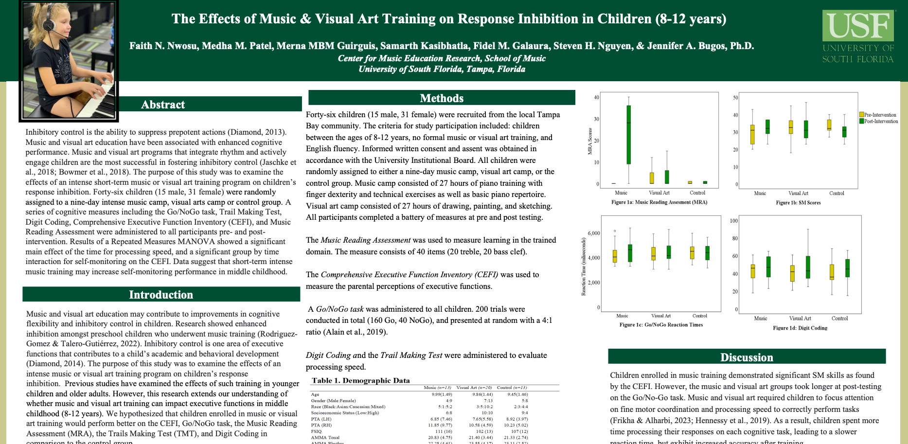
Gut Motility, Microbiota, and Neurological Disorders
Sandra Aitcheikh (sandraaitcheikh@usf.edu), Jomana Shenouda (Jomanashenouda@usf.edu), Marolina Binyamen (marolinab@usf.edu), Sydney Rivera (rivera418@usf.edu), Nikhil Chainani (nikhilchainani@usf.edu)
University of South Florida College of Public Health
Gut microbiota is the collection of microorganisms, such as bacteria and fungi, which live within human beings' digestive tract and intestines. The Gut-Brain Axis coordinates the physiological information transfer of signaling events between two sites. Any imbalances in the hormone-secreting gut can cross the blood-brain barrier and increase the dysregulation in neurological systems. The communication between the gut microbiota and the brain impacts neurological health through immune system modulation, neurotransmitter production, and vagus nerve signaling. Gut motility is the movement and stretching of the muscles in the digestive tract that allows food movement through the gastrointestinal tract. Dysbiosis is the disruption of the balance of the gut microbiota, which reduces the communication to the brain that can go through the vagus nerve. Dysbiosis has been connected to various neurological disorders such as Parkinson’s disease, Alzheimer’s disease, and mood disorders, highlighting the therapeutic potential of targeting gut health. These are all degenerative neurological diseases that are exacerbated by miscommunication between the gut and neurotransmitters. Emerging evidence suggests that gut microbiota modulate neurological processes through mechanisms including metabolite production, immune system modulation, and neural signaling. Aid to the gut can be affected using probiotics and any uncultured products. Further investigation into diet-medical interventions would be vital to reducing gut inflammation and thus provide support for healthy signaling in the gut-brain axis. This research is based on comprehensive literature reviews of peer-reviewed journal articles and scientific studies investigating the relationship between gut microbiota and neurological disorders. The data studies the link between the two in controlled experiments and through nervous and gut pathways analysis.
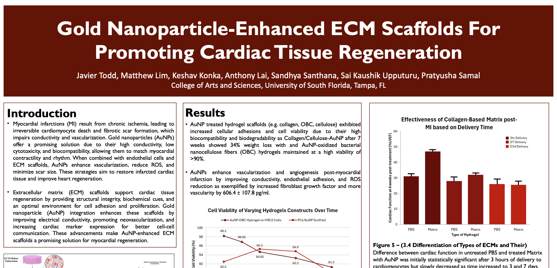
Investigation into the Correlation between Signal Transducer and Activator of Transcription 3 (STAT3) Pathways and the Development of Glaucoma
Lori Nguyen (lhnguyen285@usf.edu), Ada Lau (adalau@usf.edu), Kadence Relente (kcrelente@usf.edu), Steven Nguyen (nguyens12@usf.edu), Teerth Pansuria (teerthpansuria@usf.edu).
University of South Florida College of Arts and Science
Signal transducer and activator of transcription 3 (STAT3) is a transcriptional regulator that plays a role in the control of immunity and inflammation. Glaucoma is the degeneration of retinal ganglion cells due to high intraocular pressure, resulting in vision loss. The objective of this literature review is to consolidate existing studies regarding STAT3 and glaucoma to identify common pathways and cell regulatory processes that respond to glaucomatous injuries. A comprehensive review of 30 peer-reviewed articles with a publication range from 2004 to 2024 was conducted via PubMed, focusing on the terms STAT3, glaucoma, intraocular pressure, retinal ganglion cells, and cell signalling. A study by Wiggs (2015) found that glaucoma can be caused by mutations in proteins regulating cell division, cytokine signaling, and ocular development. STAT3 is a major signaling pathway that is involved in the regulation of cell division and retinal ganglion or axon regeneration. If STAT3 is inactivated in the optic nerve, axon regeneration is inhibited contributing to retinal ganglion cells degenerating. If STAT3 is hyperactivated in the optic nerve, cytokines can become inflamed contributing to increased intraocular pressure. Therefore, the literature suggests a relationship between the signaling pathway of STAT3 and the pathogenesis of glaucoma. Since glaucoma inhibits crucial cytokines involved with STAT3 pathways, the regenerative and healing properties of STAT3 on retinal ganglion cells are greatly reduced. Future research should be oriented towards further cross-examinations of STAT3’s impact on cellular regeneration and degeneration in glaucoma.
Stem Cell-Based Treatments for Spinal Cord Injuries
Fahed Hamad - fhamad@usf.edu
Varsha Pondicherry - varshapondicherry@usf.edu
Vaishnavi Kota - kota15@usf.edu
University of South Florida College of Arts & Sciences
Spinal cord injuries disrupt the human body’s sensorimotor functions. However, with studies on
regenerative medicine, stem cell treatments have been implemented in the central nervous
system (CNS) to restore functions after spinal cord injuries (SCI). This systematic review aimed
to assess the effectiveness of different stem cell therapies against SCI. By using “spinal cord
injuries,” “stem cell research,” and their Medical Subject Headings as its search terms, this
review yielded 370 results from PubMed. Using Rayyan, this review included ten English,
original, peer-reviewed studies between 2014 and 2024, while it excluded review articles or
studies that do not focus on human adult stem cells and SCI. Through immunosuppressive drugs,
phase I/II clinical trials confirmed stem cell therapy’s safety. From there, stem cell therapy’s
effectiveness was assessed using the American Spinal Injury Association’s Impairment Scale
(AIS). Many types of stem cells showed promising results: adipose-derived stem cells grown
alongside endothelial cells significantly enhanced neural growth with capillary recruitment.
Additionally, nearly half of the patients receiving autologous bone marrow mesenchymal stem
cells (MSC) had improved ASI grading, sensorimotor functions, and bladder usage.
Nevertheless, some stem cells yielded conflicting results. Although CNS stem cells in the
thoracic spinal cord improved senses, they did not affect the lower extremities’ motor scores.
Similarly, umbilical cord MSC significantly improved sensorimotor functions, but another study
found the lower extremities’ improved strength to be insignificant. Despite stem cell therapy’s
promising results for resolving SCI, more research is needed to improve its effectiveness.
Impact of Diabetic Retinopathy Severity on the Progression of Open-Angle Glaucoma
Esha Haque eshahaq628@gmail.com, Anvitha Makkena anvi.makkena@gmail.com, Trisha Pitchala pitchalat@usf.edu
University of South Florida of Public Health
Diabetic retinopathy (DR) and primary open-angle glaucoma (POAG) are common ocular
conditions that frequently coexist, contributing to preventable blindness. This systematic review
explores the relationship between DR severity and POAG progression, focusing on their shared
pathophysiological mechanisms. DR, a microvascular complication of diabetes, progresses
through stages classified by the ETDRS scale, with factors such as hyperglycemia,
hypertension, and diabetes duration accelerating microvascular and retinal damage. POAG, a
chronic optic neuropathy, is characterized by progressive retinal nerve fiber layer (RNFL)
thinning and is often linked to elevated intraocular pressure (IOP) and visual field deterioration.
The review synthesizes evidence from PubMed, Scopus, and other sources, highlighting how
advanced stages of DR may exacerbate POAG progression. Shared mechanisms, including
IOP fluctuations, retinal blood flow changes, vascular dysfunction, and optic nerve head
alterations, contribute to the worsening of both conditions. Understanding the link between DR
and POAG is crucial for early detection and intervention. This review emphasizes the need for
integrated management approaches, improved diagnostic strategies, and timely treatment to
prevent further ocular complications. By addressing shared risk factors, healthcare providers
can help reduce preventable blindness and improve patient outcomes globally.
Perioperative infection control and antibiotic prophylaxis for ventriculoperitoneal shunt insertion
Andres Coll andrescollsimoes@usf.edu, Luis Diaz Diaz484@usf.edu, Ana Zurita anazurita@usf.edu, Maxx Struhs maxxstruhs@usf.edu, Aishwarya Aggarwal aggarwal1@usf.edu
University of South Florida College of Arts and Sciences
Infection associated with ventriculoperitoneal (VP) shunt insertion is a problem faced by many hydrocephalus patients, often caused by gram-positive bacteria. Research has identified infection rates in neonatal and pediatric patients, and frequent procedural errors that raise the risk of infection; but the effectiveness of specific antibiotic treatments and protocols requires further exploration. This study evaluates antibiotic prophylaxis and perioperative infection control to determine the most effective preventative measures in reducing the infection rate in VP shunt insertion procedures by analyzing existing literature and comparing infection outcomes in patients. A literature review was conducted to examine the success of perioperative procedures implemented for VP shunt insertion among neonatal and pediatric patients. A total of 80 articles from PubMed were screened and 35 were selected for detailed analysis based on relevance, success rate, patient outcome, and infection incidence rates. Findings suggest that infections remain the primary complication of VP shunt insertion, but standardized protocols, antibiotics, and perioperative procedures can significantly reduce infection rates. Cephalosporin prophylaxis has been particularly effective, with a lower infection rate (3.8%) compared to cloxacillin (12.7% infection rate), due to its broad-spectrum activity against gram-positive bacteria. Patients undergoing revision surgeries experience a higher infection rate (4.8%) due to increased exposure, allowing bacteria to colonize open shunts. Cephalosporin targets Staphylococcusaureus, while cloxacillin targets Staphylococcus epidermidis, providing antimicrobial activity against these pathogens responsible for shunt-related infections. Implementing effective antibiotic treatments and protocols in insertion surgeries is crucial in reducing complications and improving long-term patient outcomes.
Implications of Ozempic: A Semaglutide with Peptide (GLP)-1 Receptor Agonists Misused for Cosmetic Weight Loss
Renee Reardon, reneeellareardon@usf.edu
University of South Florida
Ozempic, classified as a GLP-1 receptor agonist, is a semaglutide primarily prescribed for managing type 2 diabetes. It operates by mimicking the hormone glucagon-like peptide-1 (GLP-1), thereby prompting insulin secretion and reducing glucagon levels. Administered via weekly injections, it aids in regulating blood sugar levels by enhancing insulin production from the pancreas. Originally designed to assist diabetics in controlling their blood sugar, it was later discovered to be effective in promoting weight loss as well. However, individuals using Ozempic for weight loss may face elevated risks of experiencing gastrointestinal issues. This conclusion stems from comprehensive research analyzing insurance claims spanning from 2006 to 2020, involving over 5,000 patients across the United States. The findings highlighted a range of health complications, including biliary disease, gastroparesis, pancreatitis, and bowel obstructions among those using Ozempic for weight loss. Notably, collected data has revealed a significant demand for Ozempic for weight loss, with 22% of Americans seeking prescriptions from their physicians and 15% having personally used it for weight management. Additionally, 47% of respondents reported knowing someone who had used Ozempic for similar purposes. These statistics underscore the imperative for healthcare providers to educate patients regarding appropriate medication use and associated risks. Moreover, the study underscores the necessity for further research initiatives and public awareness campaigns to address the hazards associated with the misuse of pharmaceuticals for off-label purposes for cosmetic usage.
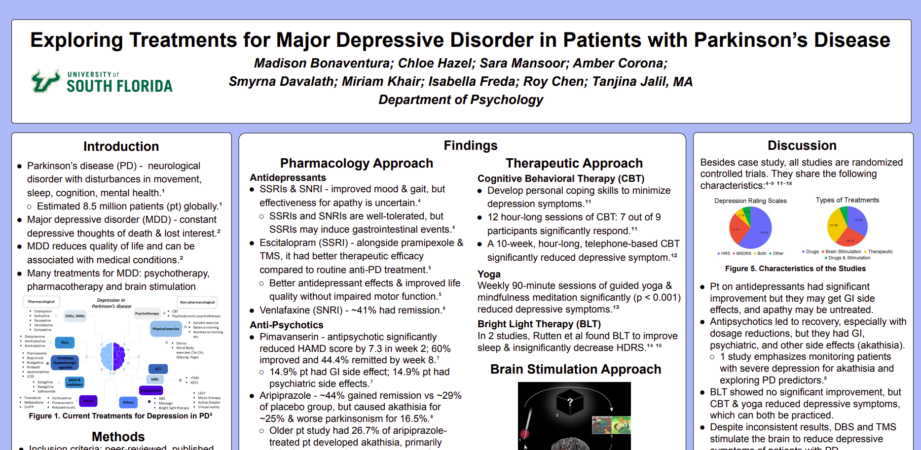
Premature Aging Biomarkers in Schizophrenia: A Correlative Relationship
Nadia Rohani; nadiar@usf.edu, Rania Jamal; jamalr@usf.edu
University of South Florida College of Arts and Sciences
Premature aging processes, characterized by biomarkers such as telomere attrition, oxidative stress, and inflammatory responses, have been increasingly recognized in individuals with schizophrenia. Previous studies suggest a possible link between these aging biomarkers and the progression of schizophrenia, yet the precise nature of this relationship remains unclear. This review aims to synthesize recent findings on aging biomarkers in schizophrenia to establish a stronger correlation between premature aging biology and schizophrenia progression, and identify potential areas for therapeutic intervention. A systematic review was conducted, analyzing 30 articles published within the past 10 years from databases including PubMed and ScienceDirect. Keywords used in the search included “schizophrenia,” “premature aging,” “telomere length,” and “oxidative stress.” The review found that individuals with schizophrenia exhibit significantly shorter telomere lengths compared to control groups, indicating accelerated cellular aging. Elevated levels of oxidative stress markers and systemic inflammation were also consistently observed among patients with schizophrenia. These biomarkers were associated with increased cognitive decline and functional impairments, suggesting a link between premature aging processes and disease severity. These findings underscore a potential mechanistic link between premature aging biology and schizophrenia progression. However, a gap remains in understanding the precise role these biomarkers play in disease development, highlighting the need for longitudinal studies. Addressing this association could inform the development of therapeutic strategies targeting aging biomarkers, potentially slowing cognitive decline and improving patient outcomes.
Reducing ICU Mortality in Sepsis Patients
Lauren Bage, Megha Kalia, Gabriella Romero-Leyva, Nivy Chandrakanth
University of South Florida College of Arts and Sciences
Sepsis is a life-threatening condition caused by an abnormal immune response, leading to excessive inflammation, multi-organ failure, and is the primary etiology of mortality in intensive care units. This review aims to interpret the effect of therapies and early detection on patient outcomes and sepsis-related mortality in the ICU. A systematic literature review was conducted using PubMed to analyze articles relevant to strategies for reducing ICU mortality published between 2023 and 2025. The review focused on 76 articles, with selected studies emphasizing vasopressor use reduction, prevention of bloodstream infections, and improvements in clinical protocols. Hypotension is a common adverse reaction to sepsis, it’s treated by vasopressors which must be minimized to reduce blood flow constriction and dependency. HAT therapy–a combination of ascorbic acid, hydrocortisone, thiamine–and methylene blue are recognized for reducing vasopressor usage and stabilizing organ function by inhibiting nitric oxide pathways to allow an increase in vascular tone. Protocols, such as the Princess Code, focus on rapid response with early recognition and intervention within one hour of diagnosis. Implementing a Comprehensive Unit-based Safety Program (CUSP) model has been associated with a 36% reduction in central line-associated bloodstream (CLABSI) rates and reduced ICU mortality. Findings from ICU mortality reviews demonstrate their critical role in improving patient outcomes through targeted interventions. Reducing vasopressor use, early detection of bloodstream infections, and improved infection protocols contribute to better hemodynamic stability, rapid diagnostics, and antimicrobial stewardship. Standardized ICU protocols are essential in reducing sepsis-related mortality, reinforcing the need for their widespread implementation.
Provider Engagement in ICU Quality and Process Improvement
Sailaasya Pinnamaneni (sailaasyap@usf.edu), Nour Shokr (nourshokr@usf.edu), Sama Shokr (shokr@usf.edu), Carole Wadie (carolew@usf.edu), Niveditha Chandrakanth (nchandrakanth@usf.edu)
University of South Florida College of Arts and Sciences
Effective provider engagement in the Intensive Care Unit (ICU) is critical for optimizing patient outcomes and ensuring high-quality care delivery. This study examines the relationship between provider engagement and ICU process improvements. Family/patient-centered care, interprofessional collaboration, and education/training are key factors to enhance engagement, fostering a more efficient care environment. A systematic literature review was conducted using PubMed, incorporating quantitative and qualitative data to understand provider engagement and processes in the ICU. Of the 77 articles screened, 24 met the criteria. Shared decision-making between the provider and the patient/family led to a 29% reduction in adverse events and a significant increase in patient satisfaction from 71.8 to 93.3. Participation in rounds potentially enhances outcomes in the ICU. Collaborative practice has improved patient outcomes, leading to a 43% reduction in ICU mortality and a decrease in ICU length of stay from 8.3 to 3.6 days. An essential element was the continuous education and training of the providers; in particular, case-based learning and on-the-job training were the most impactful in modifying established processes. These aspects improve ICU processes by reducing complications, such as a 69% decrease in CLABSI rates in one year, through a holistic approach to care. This review provides a structured approach to how high-quality care in the ICU can be enhanced and further assessed through interviews and surveys with healthcare professionals, patient families, and ICU patients. Future studies should integrate longitudinal data alongside critical provider feedback to enhance ICU care.
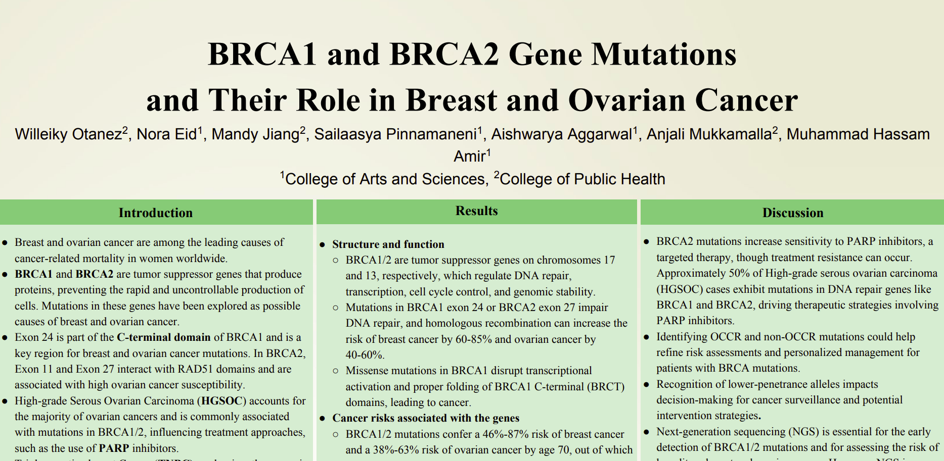
Epigenetic Modifications that cause Type 1 Diabetes: Exploring DNA methylation and Histone changes in Autoimmune Pathogenesis
Sebastian Tirado
satirado@usf.edu
University of South Florida College of arts and sciences GHC
Type one diabetes is an autoimmune disease that is characterized by the destruction of beta cells in the pancreas. Genetic predisposition plays a major role in the development of Type 1 Diabetes, new data suggest that epigenetic modifications such as DNA methylation and histone modifications contribute to disease onset and progression. This literature review analyzes a variety of recent epigenomic-wide association studies to identify modifications leading to the development in Type 1 Diabetes. Studies examining DNA methylation patterns in immune pancreatic cells, the impact of environmental factors on the epigenome, and histone modifications affecting inflammatory pathways were analyzed. Findings showed that hypermethylation of immune-related genes and hypomethylation of regulatory T-cell genes disrupted immune tolerance. Histone modifications, such as altering H3K27 acetylation, have been linked to pro-inflammatory gene expression in beta cells (insulin producing cells). Additionally, many environmental factors such as diet or viral infections appear to aggravate these epigenetic changes which increase development for type one diabetes. Epigenetic modifications, (genetic or environmental) contribute a major role in Type 1 diabetes pathogenesis. Further research can be used to develop targeted epigenetic therapies to help preserve beta cell function or modify immune system responses under these conditions.

