Optimizing Neuro Check Frequency to Prevent Early Neurological Deterioration in Stroke Patients: A Systematic Review
Krutika Pedamkar (kspedamkar@usf.edu), Joseph Tobin (jrtobin@usf.edu), Jasmin Salcedo-Lara (jasminsalcedolara@usf.edu), Raneem Yassin (raneemyassin@usf.edu) , Aishwarya Aggarwal (aggarwal1@usf.edu)
University of South Florida College of Arts and Sciences
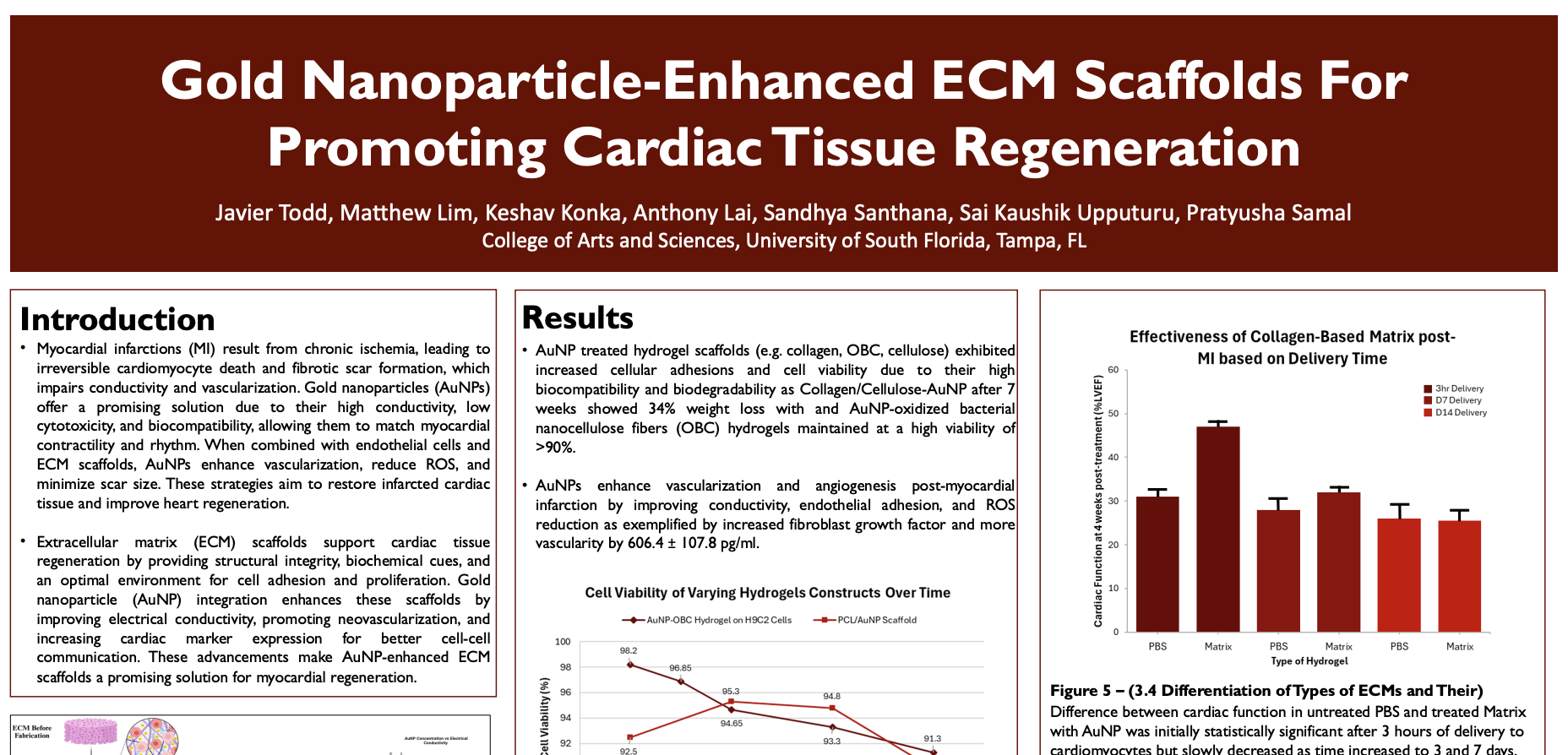
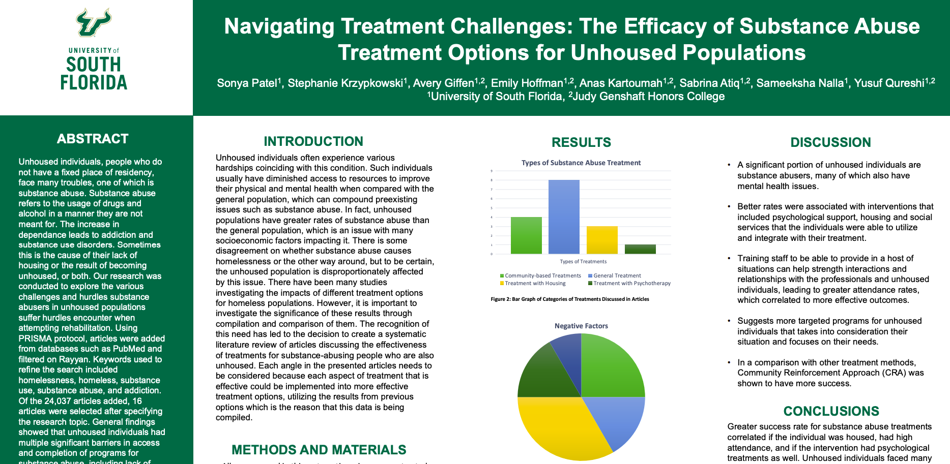
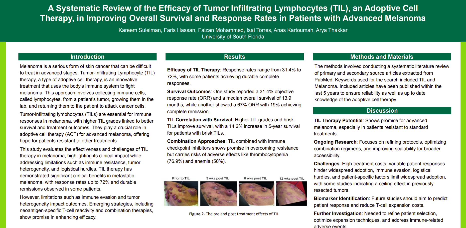
Neuro checks are essential for detecting early neurological deterioration (END) in ischemic and hemorrhagic stroke patients, especially during the first 24 hours. While frequent assessments are critical in this early phase, research suggests that their effectiveness diminishes beyond this period. Current protocols have limitations, particularly in detecting gradual or subtle changes. The potential consequences of prolonged neuro checks, like sleep disruption or delirium, highlight the need for a balanced approach to monitoring in stroke care. This study examines the impact of neuro check frequency on the detection of evolving END in stroke patients. It also identifies optimal monitoring strategies for improving detection and patient outcomes. A comprehensive review of 32 peer-reviewed articles (2014-2024) from PubMed examined neuro check frequency in acute stroke patients, END detection, and clinical outcomes. Studies lacking patient monitoring data or insufficient sample sizes were excluded. Findings indicate that approximately 27% of stroke or neurologic disease patients experience END. Nearly half of these cases are detected through scheduled neuro checks. A high frequency of neuro checks (hourly) increases the likelihood of early detection; however, their effectiveness declines beyond the first 12–24 hours due to reduced detection rates of new deterioration, as most critical neurological changes occur within the first day. Prolonged neuro checks, especially beyond 24 hours, are linked to a threefold increase in delirium risk due to sleep disruption. Adjusting or discontinuing checks after 12–24 hours can mitigate these risks, while alternative monitoring strategies can ensure early detection of neurological deterioration.
The Effects of Microplastic Phytotoxicity on Seagrass and Duckweed
Wynter R. Dean wynterrd7@gmail.com, Coen E.E. McGarrah mcgarrah@usf.edu, Brenna Strehle strehle.brenna@gmail.com, Aurora Gomez Garcia gomezgarcia@usf.edu, Huong Ha Lan Do lhdo@usf.edu, Katelynn Paciorek katelynnpaciorek@usf.edu
University of South Florida College of Arts and Sciences
Microplastics are plastic particles ranging from one nanometer to five millimeters and are comprised of various chemical compounds. These particles are thought to have wide health and environmental impacts, including restricted growth and photosynthesis of aquatic plants such as duckweeds and seagrasses. These plants are essential for maintaining marine ecosystems, improving water quality, and protecting the coast. This review compiles current knowledge on the abundance and biological effects of microplastic interactions with aquatic plants, using duckweed and seagrass as model organisms. In initial searches for relevant literature, approximately 2,000 articles were selected for review based on their titles and uploaded to EndNote, where duplicate articles were eliminated. The selected articles were then transferred to Rayyan for full review of the papers’ contents, with 39 papers included in the final review. The precise mechanism of microplastic phytotoxicity on seagrasses and duckweeds has yet to be thoroughly investigated, though general effects have been observed. Studies on existing populations are limited. Plant growth impairment appears to vary depending on microplastic type. Two common effects are impairment of seagrass blade or duckweed root growth, and cytoskeletal defects. Small particles could be absorbed directly into plant tissues and coat their exteriors. Additionally, stress from microplastics can interact with other aquatic plant contaminants. Impacts on primary consumers have been noted in both species contaminated with microplastics. From these findings it is clear that microplastics have a negative effect on aquatic plants, though more research is needed to determine the extent of their impact.
GPCR-Mediated Mechanisms in Obesity: The Role of Ghrelin and GLP-1 Receptors
Ryan Singh (rs49@usf.edu), Tuana Cenberoglu (tuanac@usf.edu),
Jonathan Budlong (jbudlong@usf.edu),
Jaclyn Alejandro (jaclynrachel@usf.edu), and
Andrea Weitoschova
University of South Florida College of Arts and Sciences
G protein-coupled receptors (GPCRs) regulate metabolism and transmit appetite cues throughout the brain and digestive system. Binding to GPCRs, ghrelin, and GLP-1 are ligands that influence hunger, energy balance, and insulin secretion. Understanding the relationship between GPCRs and ligands such as ghrelin and GLP-1 offers critical insights into treating metabolic disorders such as obesity, a growing issue in America. Our methodology involved a literature review using USF, NIH, and JSTOR databases. Search terms like “GPCR obesity,” “GLP-1 obesity,” and related keywords identified articles from 2000 onward, yielding 59 studies on GLP-1 and ghrelin in obesity. GLP-1 receptor agonists (GLP-1RAs), initially developed for type 2 diabetes, have shown effectiveness in treating obesity by reducing blood glucose levels, appetite, and hunger while delaying gastric emptying and enhancing satiety. Studies on semaglutide, liraglutide, and retatrutide demonstrated their efficacy in promoting weight loss when combined with diet and exercise, highlighting their potential as pharmacologic treatments for obesity. However, side effects such as gastrointestinal discomfort and limitations of mostly animal studies underscore the need for further research to enhance safety and reliability. This study highlights the pivotal role of GPCRs in managing obesity and diabetes by regulating energy balance and metabolism. Advances in GLP-1-based therapies and GPCR-targeting drugs offer transformative potential for personalized treatments, emphasizing the importance of continued research into these mechanisms.
GCPR’s Roles in Inflammatory Bowel Disease
Lorenzo Macatol @lorenzomacatol@usf.edu
Aurora Gomez gomezgarcia@usf.edu
Allha Akrami - aakrami@usf.edu
Reanna clean mclean350@usf.edu
University of south Florida of arts and sciences
GPCRs, or g-reactive protein receptors, are signaling proteins that possess a significant impact on the immune and inflammatory responses of IBD patients. Past research has discovered the relationship of GPCRs and its regulation of inflammation in mediated pathways. However, this literature review achieves a deeper understanding of this regulation involving the impacts of the bacterial microbiome and immune system of the intestinal barrier on macrophage and GPCR-mediated pathways. The aim of this literature review is to gain knowledge and clarity in the utilization of GPCRs in IBD. This literature review discovers relationships between these proteins and the intestinal barrier, inflammation control, and immune dysfunction of IBD patients. Method
This study conducted a literature review of 30 peer-reviewed articles examining the roles of GPCRs in IBD pathogenesis, inflammation regulation, and therapeutic potential. Articles were sourced from databases including Google Scholar and the USF Library, focusing on recent publications to ensure relevance. The analysis synthesized data on GPCR functionality, SCFA interactions, and their implications for intestinal homeostasis and inflammation control.
Results:
Our findings highlight the critical role of the immune system and bacterial microbiome in regulating inflammatory responses through macrophage and GPCR-mediated pathways. Notable GPCRs, including GPR43, GPR109A, and GPR35, contribute to maintaining intestinal barrier integrity and inflammation control, with SCFA-GPCR interactions promoting immune regulation and gut homeostasis. Additionally, GPCRs such as GPR183 and GPR40 demonstrate therapeutic potential by supporting mucosal regeneration and alleviating inflammatory bowel disease (IBD) symptoms. These insights underscore the pharmaceutical relevance of GPCR modulation in treating inflammation-driven intestinal disorders.
Conclusion Current forays into the relationship between GCPRs and IBD reveal consiste
Socioeconomic Disparities and Disruption of Circadian Gene Expression in PMBCs: Implications for Chronic Disease Risk
Bhaveshsai Reddy bhaveshsaireddy@usf.edu, Eshan Baig eshan4510@gmail.com, Dev Thakkar Devthakkar@usf.edu, Ryan Chowdhury chowdhury60@usf.edu
University of South Florida College of Arts and Sciences
This study explores the relationship between relative poverty and the regulation of genes related to circadian rhythm in peripheral blood mononuclear cells (PBMCs), focusing on how socioeconomic factors may correlate with disrupted biological circadian rhythms and subsequent health outcomes. Disruptions in circadian rhythm are a catalyst for many adverse downstream effects: impaired immune function, increased inflammation, and most concerningly, elevated risk of developing chronic diseases. Understanding gene expression in PBMCs specifically is key in studying circadian regulation as it provides a model for systemic immune function, which is influenced by circadian rhythms. We investigate the relationship between socioeconomic status and circadian gene expression in PBMCs using data from a cross-sectional study of older persons with and without a history of depression in various socioeconomic circumstances. The scope of our research is specific to genes involved in immune response, inflammation, and stress adaptation (CLOCK, BMAL1, and PER2). Our initial findings suggest that individuals living in poverty exhibit significant disruptions in circadian gene expression correlated to socioeconomic gradients. The working hypothesis is that disruptions can be further linked to higher levels of systemic inflammation and increased risk of chronic diseases, with a focus on cardiovascular conditions. The stated goal of our study is then to explore targeted interventions that could be aimed at restoring circadian rhythm function in individuals affected by poverty, which may improve overall health outcomes and reduce health disparities through a basic understanding of the molecular mechanisms that have been studied.
Environmentally Adaptive Prosthetic Foot: Enhancing Mobility and Stability with Bioinspired Soft Robotics
Yara Saleh saleh21@usf.edu, Maxwell Weisel maxwellweisel@usf.edu, Dhruvi Patel dhruvip@usf.edu, Dontavious Ellis Dellis@usf.edu, Kieran Ball Ballk141@usf.edu
University of South Florida College of Engineering
Traditional prosthetic feet struggle to adapt to varying terrains, limiting mobility and increasing fall risk for amputees. This research explores the development of an environmentally adaptive prosthetic foot, influenced by bioinspired soft robotics, to enhance stability and energy efficiency across diverse surfaces. Using a biomimetic design similar to the SoftFoot Pro (CYBATHLON, 2025), the prosthetic features a flexible and segmented structure that dynamically adjusts for stiffness. Additionally, a shock-absorbing mechanism at the heel is designed to enhance energy return and stability by reducing the impact forces and improving adaptability during ground interactions. This design provides improved balance and support across various terrains. A finite element analysis will be performed to assess the prosthetic's response to stress under different forces. Currently, the initial design is in the troubleshooting phase to refine the functionality and ensure optimal performance. Challenges related to material durability, structural integrity, and adaptive responsiveness are being addressed through constant repetitive prototyping and mechanical testing. By leveraging soft robotics and a shock-absorbing mechanism, this approach is expected to enhance stability and energy efficiency by offering a more natural and responsive experience for users. This advancement pushes prosthetic technology toward greater functionality and comfort by improving adaptability to varied terrains. Future work will focus on refining the experimental gait analysis of the shock-absorbing mechanism and expanding field testing to optimize long-term performance in diverse environments.
Challenges and Innovations in Pediatric Anesthesiology
Dana John (danajohn@usf.edu), Khushi Jariwala (khushijariwala@usf.edu), Delilah Penate (dpenate@ufl.edu), Emma Chacko (echacko@usf.edu), Anandita Makkar (ananditamakkar@usf.edu), Stuti Dibbur (stutid@usf.edu)
University of South Florida College of arts and sciences
Background: Pediatric anesthesiology is a field that must account for the physiological and drug metabolic differences between children and adults. Previous studies acknowledge the challenges posed by pediatric airway management and brain health risk. This literature review aims to address advances in pediatric airway management via videolaryngoscopy and brain health innovation via near infrared spectroscopy in the field of pediatric anesthesiology.
Methods: This study evaluated research on key technological advancements in NIRS and videolaryngoscopy to examine promising advances for the related persistent challenges in pediatric anesthesiology.
Results: The literature suggests that advances in the use of NIRS, can measure cerebral oxygenation and lower neurologic injury risk. Low cerebral oxygenation levels after a cardiac surgery have been linked to poor neurodevelopmental outcomes, thus making NIRS an effective tool for anesthesiologists to monitor and adjust factors such as ventilation, perfusion, or blood pressure as needed. The more routine use of videolaryngoscopy has also provided a potential method of combating uncertainties in navigating pediatric airways, particularly when combining two video enabled devices—a laryngoscope and a fiberscope. This advancement provides a live visual to ensure proper and less traumatic placement of the endotracheal tube, reducing esophageal intubations and airway trauma.
Discussion/Conclusion: The ability to manage and care for children continues to evolve, and the ability to assess tissue oxygenation of tissues deep to the skin and to more reliably and easily access and control pediatric airways represent two noteworthy advances for patients and the providers who care for them.
GPCRs in Rheumatoid Arthritis: Role of Chemokine Receptors in Joint Inflammation
Anthony Deepesh deepeshanthony@gmail.com, Umesh Mohann umeshchandramohann@gmail.com, Willeiky Otanez willeikyod@gmail.com, Harinandan Nisheed harinandan.nisheed@gmail.com, Andrea Weitoschova a.weitoschova@gmail.com
University of South Florida
Rheumatoid arthritis (RA) is a chronic autoimmune disease characterized by joint inflammation and damage. G protein-coupled receptors (GPCRs), particularly chemokine receptors, play a crucial role in recruiting and activating immune cells in the inflamed synovium, contributing to RA pathogenesis. This study aims to elucidate the involvement of GPCRs in RA’s inflammatory processes, highlighting their significance in disease mechanisms and potential as therapeutic targets. A systematic literature review was conducted using PubMed, employing keywords such as “GPCR,” “chemokine receptors,” “rheumatoid arthritis,” and “inflammation,” which led to the identification of 67 relevant articles from the past two decades that assess the role of GPCRs and chemokine receptors in RA. The review found that chemokines and their receptors, such as CCR1, CXCR3, and CCR2, are significant in RA pathogenesis by promoting leukocyte recruitment, retention, and inflammatory responses in synovial tissues. Targeting these chemokine receptors has shown therapeutic benefits in animal models, reducing joint inflammation and damage. However, clinical trials in humans have yielded mixed results due to pathway redundancies and regulatory complexities. Findings suggest that combination therapies targeting multiple chemokine receptors could enhance treatment efficacy by blocking compensatory mechanisms. This study underscores the critical role of chemokine receptors in RA pathogenesis and their therapeutic potential despite challenges with pathway redundancies. Limitations include translational barriers; further research should explore synergistic receptor-targeting strategies to improve therapeutic specificity and efficacy. GPCRs and chemokine receptors remain pivotal in RA by driving inflammation and immune cell recruitment. Advancing combination therapies and receptor-specific research may enhance therapeutic outcomes.
What is the role of aberrant tyrosinase in vitiligo in pediatric versus geriatric patients?
Joshua Robert Diaz - diaz241@usf.edu
University of South Florida College of Arts and Sciences
This research examines whether a digital platform, Tsugi, employing AI-driven peer mentorship, can reduce mental health stigma and improve access to mental health support, thereby decreasing rates of depression and anxiety among amputees. Amputees face disproportionately high rates of mental health challenges, with 35% experiencing depression and 60% suffering from anxiety within the first six months after limb loss (Jo et al., 2021). Stigma surrounding both limb loss and mental health exacerbates these challenges, discouraging individuals from seeking help. Existing peer-support programs, such as AMPOWER and the Amputee Coalition app, struggle with extreme volunteer dependency and limited personalization, reducing their effectiveness (Amputee Coalition, 2024; Clean with Ross, 2024). This study explores the potential of Tsugi, an AI-driven digital mentorship platform, to address these limitations by fostering a stigma-free support environment through peer mentorship. Preliminary findings suggest that structured digital mentorship significantly improves self-perception, reduces social anxiety, and enhances help-seeking behaviors within the prosthetic community (Gainesville Prosthetics, 2023). Additionally, 75% of amputees report that peer support positively impacts their outlook on life, while 78% find the shared information beneficial (Lee et al., 2024). By leveraging AI-integrated digital ecosystems, like Tsugi, this research aims to dismantle deeply ingrained stigmas and establish scalable, technology-driven psychological support models for amputees worldwide. Addressing both emotional and practical challenges, Tsugi fosters a more inclusive and empowering recovery experience for amputees, ensuring long-term engagement with mental health care.
MORTALITY AND INCIDENCE DISPARITIES IN BREAST CANCER: SOCIOECONOMIC INFLUENCES ACROSS ETHNIC GROUPS
Merna Guirguis guirguis3@usf.edu, Christiana Mihaila christianamihaila@usf.edu, Carla Gutierrez-Sotelo cgutierrezsotelo@usf.edu, Daniela Lozada danielaburgalozada@usf.edu, Jun Zheng junjiezheng@usf.edu, Yen Lee hoang11@usf.edu, JM Warith Rahman jmrahman@usf.edu
University of South Florida College of Arts and Sciences
Breast cancer is the second leading cause of cancer-related deaths among U.S. women, with over 42,000 deaths projected in 2024. Socioeconomic status (SES) significantly influences healthcare access, treatment quality, and outcomes. Disparities in SES contribute to variations in breast cancer incidence, progression, and mortality, disproportionately affecting racial and ethnic minorities. African American women experience the highest mortality rates despite advancements in treatment, largely due to financial barriers, late-stage diagnoses, and gaps in insurance coverage. A systematic review was conducted using AI-powered Elicit and databases like PubMed, NIH, and Google Scholar, analyzing studies from 2020–2025. The focus was on breast cancer outcomes in U.S. women under 30, emphasizing racial and ethnic disparities. SEER*Explorer and State Cancer Profiles provided data on incidence, mortality, and survival rates. Results indicate that African American women face disproportionately high mortality, with incidence rates between 26,000 and 28,000 cases from 2017 to 2021. Between 2018 and 2022, breast cancer deaths among non-Hispanic Black females aged 15 to 39 ranged from 3,600 to 4,000—nearly double that of non-Hispanic White females. Lower median household incomes, reduced access to preventive care, and systemic healthcare inequities contribute to delayed diagnoses and poor survival rates. Additionally, geographic disparities, implicit provider biases, and lower treatment adherence further exacerbate healthcare barriers. Addressing these disparities requires expanding healthcare access, policy interventions, and targeted community outreach programs. Future research should integrate genetic, environmental, and psychosocial factors to develop comprehensive strategies aimed at reducing disparities. By tackling systemic inequities, significant progress can be made in lowering breast cancer mortality among underserved populations and improving national health outcomes.

